Home > Images Gallery
Images Gallery
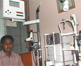
Images - Courtesy Dr. S.S. Bhatti
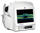
Carl Zeiss HD OCT

Heidelberg Confocal Imaging System West Germany

Sonomed Ultra Sound A and B Scan USA
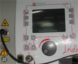
Iridis laser For Retina - Quantel medical France
Chair Unit with Tomey Auto Refracto Heter Japan and Slit Lamp Reichart Germany

Viteretomy and Phaco Emulsification Machine
Stores - USA
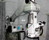
MDO operating Microscope Carlzeirs West Germany
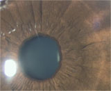
Normal iris pattern
Busaccas granulomatous
nodules on iris surface

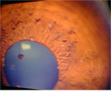
Festooned pupils due to
posterior synechiae
Peripheral anterior synechiae

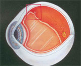
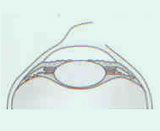
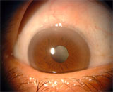
Posterior chamber IOL with uveitis, IOL capture and lens precipitates

Uveitis
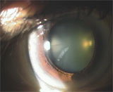
Zonular cataract

Blue dot cataract 1

Blue dot cataract 2
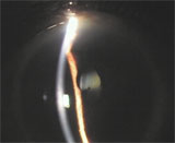
Nuclear and posterior subcapsular cataract seen on slit lamp
Vacuoles in lens fibres 1
(incipient cortical cataract)

Vacuoles in lens fibres 2
(incipient cortical cataract)
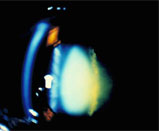
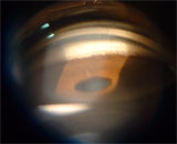
Posterior subcapsular cataract
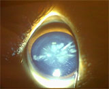
Traumatic rosette cataract
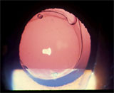
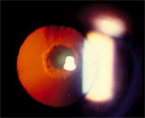
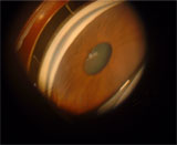
Posterior chamber IOL seen against red glow

Posterior Chamber IOL seen against the red fundus glow
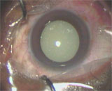
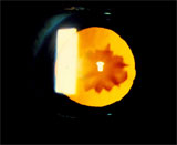
Hypermat cataract

Kelman Multiflex anterior chamber IOL
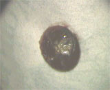
Nucleus of brown cataract
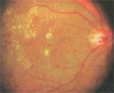
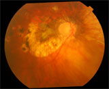
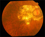
Macula is very wet and swollen (a great amount of fluid and exudate) 1

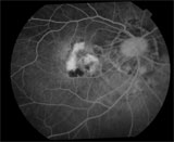
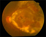
Macula is now dry 2
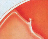
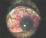
Rubeosis (neovascularization on iris)

Macula is normal and vision is normal
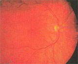
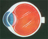
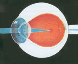
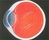
Normal Retina
optic nerve
center of retina (macula)
side (peripheral) retina

Center (macula) not treated
laser spots not in center
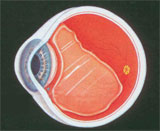
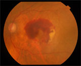
Panretinal laser photocoagulation
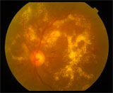
Diabetic Retinopathy
Images - Courtesy Dr. S.S. Bhatti
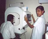
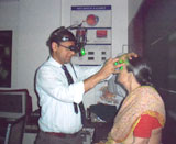
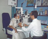
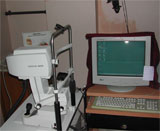
Images of Clinic and equipment
Carl Zeiss HD OCT
Heidelberg Confocal Imaging System West Germany
Sonomed Ultra Sound A and B Scan USA
Iridis laser For Retina - Quantel medical France
Chair Unit with Tomey Auto Refracto Heter Japan and Slit Lamp Reichart Germany
Viteretomy and Phaco Emulsification Machine
Stores - USA
MDO operating Microscope Carlzeirs West Germany

Normal iris pattern
Busaccas granulomatous
nodules on iris surface
Festooned pupils due to
posterior synechiae
Peripheral anterior synechiae
Posterior chamber IOL with uveitis, IOL capture and lens precipitates
Uveitis

Zonular cataract
Blue dot cataract 1
Blue dot cataract 2
Nuclear and posterior subcapsular cataract seen on slit lamp
Vacuoles in lens fibres 1
(incipient cortical cataract)
Vacuoles in lens fibres 2
(incipient cortical cataract)
Posterior subcapsular cataract
Traumatic rosette cataract
Posterior chamber IOL seen against red glow
Posterior Chamber IOL seen against the red fundus glow
Hypermat cataract
Kelman Multiflex anterior chamber IOL
Nucleus of brown cataract
Macula is very wet and swollen (a great amount of fluid and exudate) 1
Macula is now dry 2
Rubeosis (neovascularization on iris)
Macula is normal and vision is normal
Normal Retina
optic nerve
center of retina (macula)
side (peripheral) retina
Center (macula) not treated
laser spots not in center
Panretinal laser photocoagulation
Diabetic Retinopathy